
- Testing of muscles and nerves involved with cervicobrachial syndromeTesting of muscles and nerves involved with...
- Biceps reflex examinationBiceps reflex examination
- Triceps reflex examinationTriceps reflex examination
- Muscles and nerves involved with Cervicobrachial SyndromeMuscles and nerves involved with Cervicobrachial Syndrome
- Extensor Carpi Radialis ReflexExtensor Carpi Radialis Reflex
- Transport of T4 and T3 through the blood-brain-barrierTransport of T4 and T3 through the...
- synapssynaps
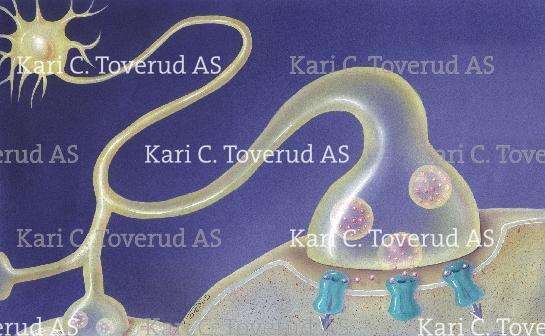
- neuromuscular junctionneuromuscular junction
- plantar reflex-Babinski´s signplantar reflex-Babinski´s sign
- Anterior Interosseous Nerve nerve injury. Cannot do the O.K signAnterior Interosseous Nerve nerve injury. Cannot do...
- Pathology of Alzheimer´s disease with tau proteins and amyloid plaquesPathology of Alzheimer´s disease with tau proteins...
- digital nerve and median nerve innervation (palmar viewdigital nerve and median nerve innervation (palmar...
- n. radialis, sensory innervationn. radialis, sensory innervation
- n. radialis, sensory innervationn. radialis, sensory innervation
- NERVE INNERVATION OF THE BLADDERNERVE INNERVATION OF THE BLADDER
- anterior view of the thyroid gland with blood blood supply and nervesanterior view of the thyroid gland with...
- lateral view of the thyroid gland with blood and nerve supplylateral view of the thyroid gland with...
- injecting novocaine into fingerinjecting novocaine into finger
- venous supply of the brainvenous supply of the brain
- cervical prolapscervical prolaps
- areas of pain from dermatomes C5,6,7 and 8areas of pain from dermatomes C5,6,7 and...
- LATERAL VIEW OF BRAIN WITH MAJOR SENSORY, MOTOR AND ASSOCIATION AREAS OF COTEXLATERAL VIEW OF BRAIN WITH MAJOR SENSORY,...
- Brachial plexusBrachial plexus
- the limbic systemthe limbic system
- prolapsed disc and recurrent meningeal nerves, sinuvertebral nerves, or recurrent nerves of Luschkaprolapsed disc and recurrent meningeal nerves, sinuvertebral...
- interlaminar and transforaminal Epidural injection in with herniated diskinterlaminar and transforaminal Epidural injection in with...
- epidural injection in sacral hiatus with herniated disk S2epidural injection in sacral hiatus with herniated...
- Caudal epidural injection techniqueCaudal epidural injection technique
- Interlaminar and transforaminal epidural injection techniqueInterlaminar and transforaminal epidural injection technique
- lateral view of brain in a femalelateral view of brain in a female
- sagital section of brain in a femalesagital section of brain in a female
- lateral view of cerebral lobes of brainlateral view of cerebral lobes of brain
- Lateral view of brain with major sensory, motor and association areas of cotexLateral view of brain with major sensory,...
- lateral view of brain within the skull of a femalelateral view of brain within the skull...
- nerve innervation of the pelvic region of a malenerve innervation of the pelvic region of...
- sacral nerves in pelvissacral nerves in pelvis
- digital blockade of fingerdigital blockade of finger
- motor nerve cellmotor nerve cell
- Pathway for embolism to the brainPathway for embolism to the brain
- Sciatic nerve painSciatic nerve pain
- Coronal section showing normal brain on the left and a brain affected with Alzheimer�s on the rightCoronal section showing normal brain on the...
- Lateral view of the brain showing blood supplyLateral view of the brain showing blood...
- Registration of EEG (electroencephalogram) during anethesiaRegistration of EEG (electroencephalogram) during anethesia
- trigeminal neuralgiatrigeminal neuralgia
- The meninges of the brain; dura mater, arachnoid, and pia materThe meninges of the brain; dura mater,...
- subarachnoid hemorrhage / bleeding (stroke)subarachnoid hemorrhage / bleeding (stroke)
- Lateral viw of the brain showing the ventriclesLateral viw of the brain showing the...
- Lateral veiw of the brain showing major sensory, motor and association areas of the cortexLateral veiw of the brain showing major...
- Lateral view of the brain with branches of the left middle cerebral artery on the brain surface in aLateral view of the brain with branches...
- A shunt from the lateral ventricle to the peritoneal cavity to treat an infant with hydrocephalusA shunt from the lateral ventricle to...
- Circulation of cerebrolspinal fluidCirculation of cerebrolspinal fluid
- Anterior view of the circulatory system and the nervous systemAnterior view of the circulatory system and...
- Lateral veiw of the brain showing major sensory, motor and association areas of the cortexLateral veiw of the brain showing major...
- Network of myelinated and unmyelinated nerve cells joined with synapses and on nerve cell connectedNetwork of myelinated and unmyelinated nerve cells...
- The action of levodopa (orange pill) and dopamine antagonist (yellow pill) on a synaps in the basalThe action of levodopa (orange pill) and...
- Lateral veiw of the brainLateral veiw of the brain
- Position of a patient during a spinal tapPosition of a patient during a spinal...
- Cross section through the lumbar section (L1) of the spinal cordCross section through the lumbar section (L1)...
- Section of the spinal cord and spinal roots showing the 3 layers of the meningesSection of the spinal cord and spinal...
- An unmyelinated and a myelinated nerve cellAn unmyelinated and a myelinated nerve cell
- Anatomy of a nerveAnatomy of a nerve
- Network of nerve cellsNetwork of nerve cells
- A nerve cell with synaptic vesicles, a synaps on a postsynaptic dentrite showing the synaptic cleftA nerve cell with synaptic vesicles, a...
- Circulation of cerebrolspinal fluidCirculation of cerebrolspinal fluid
- Medial surface of the brain, spinal cord within the cranium in a boyMedial surface of the brain, spinal cord...
- Medial surface of the brain, spinal cord within the craniumMedial surface of the brain, spinal cord...
- Medial surface of the cerebrumMedial surface of the cerebrum
- Lateral veiw of the brain and spinal cordLateral veiw of the brain and spinal...
- Medial surface of the brain and spinal cordMedial surface of the brain and spinal...
- Frontal view of the the skull and brain showing the placement of an epidural hematomaFrontal view of the the skull and...
- Creative and logical sides of the brainCreative and logical sides of the brain
- Cross section through the forebrain at mid-thalamusCross section through the forebrain at mid-thalamus
- Lateral view of the brain showing cerebral lobesLateral view of the brain showing cerebral...
- Lateral viw of the brain showing the ventriclesLateral viw of the brain showing the...
- Lateral view of the brain showing the placement of the basal ganglionLateral view of the brain showing the...
- Lateral view of the brain showing cerebral lobesLateral view of the brain showing cerebral...
- Lateral veiw of the brainLateral veiw of the brain
- Lateral view of the brain and spinal cordLateral view of the brain and spinal...
- Anterior view of the central and peripheral nervous system and an enlargement of a peripheral nerveAnterior view of the central and peripheral...
- Lateral view of the central and peripheral nervous systemLateral view of the central and peripheral...
- Anterior view of the central and peripheral nervous systemAnterior view of the central and peripheral...
- Frontal view of the brainFrontal view of the brain
- 4 common areas where the brain tissue can herniate4 common areas where the brain tissue...
- Knee jerkKnee jerk
- Sagittal section through the spinal columnSagittal section through the spinal column
- Lateral view of 3 lumbar vertebrae showing the spinal cord and the spinal nervesLateral view of 3 lumbar vertebrae showing...
- Different types of nerve cellsDifferent types of nerve cells
- Superior view of the lumbar vertebraSuperior view of the lumbar vertebra
- Superior view of the cervical vertebraSuperior view of the cervical vertebra
- Lateral view of the brain showing different causes of a cerebral strokeLateral view of the brain showing different...
- Ventral view of the brain showing the 12 pairs of cranial nervesVentral view of the brain showing the...
- Somatotopic map of the body surface onto the primary somatosensory cortex and primary motor cortexSomatotopic map of the body surface onto...
- Lateral view of the brain and spinal cordLateral view of the brain and spinal...
- Lateral veiw of the brain showing major sensory, motor and association areas of the cortexLateral veiw of the brain showing major...
- Concussion results from a blow to the headConcussion results from a blow to the...

A nerve cell with synaptic vesicles, a synaps on a postsynaptic dentrite showing the synaptic cleft
Request information on how to purchase usage rights to this image
Please fill out the form below with all the relevant details. If want to request information on more than one illustration, you may prefer to e-mail me directly instead. If you do, please include the link to this illustration and the other illustrations: http://www.karitoverud.com/illustrations/C/349/
 Instagram