
- SAM spilt lower legSAM spilt lower leg
- SAM splint for underarmSAM splint for underarm
- RICE (rest, ice, compression, elevation)RICE (rest, ice, compression, elevation)
- how to create free airwayshow to create free airways
- ankle braceankle brace
- wrist bracewrist brace
- how to tape a sprained fingerhow to tape a sprained finger
- how to apply an ace bandage on the wristhow to apply an ace bandage on...
- How to put on a splint for underarm fractureHow to put on a splint for...
- how to put on a spring for a fractured humerushow to put on a spring for...
- ace bandage on ankleace bandage on ankle
- How to tape the ankel using an ace bandageHow to tape the ankel using an...
- how to tape the akilles tendonhow to tape the akilles tendon
- brachialis musclebrachialis muscle
- extensor pollicis brevis muscleextensor pollicis brevis muscle
- extensor pollicis longus muscleextensor pollicis longus muscle
- extensor carpi ulnaris muscleextensor carpi ulnaris muscle
- extensor carpi radialis brevis muscleextensor carpi radialis brevis muscle
- extensor carpi radialis longus muscleextensor carpi radialis longus muscle
- extensor-digitorum muscleextensor-digitorum muscle
- brachioradialis musclebrachioradialis muscle
- medial epicondyle tendinopathymedial epicondyle tendinopathy
- TorticollisTorticollis
- maseter musclemaseter muscle
- Tendon attached to bone showing a normal tendon, tendinosis and tendinitisTendon attached to bone showing a normal...
- sciatic nerve in relation to the periformis musclesciatic nerve in relation to the periformis...
- periformis syndromeperiformis syndrome
- tensor fascia latae-bursitis anterior and posterior viewtensor fascia latae-bursitis anterior and posterior view
- tensor fascia latae bursitis -anterior-posttensor fascia latae bursitis -anterior-post
- trochanter tendinitistrochanter tendinitis
- plantar reflex-Babinski´s signplantar reflex-Babinski´s sign
- Anterior Interosseous Nerve nerve injury. Cannot do the O.K signAnterior Interosseous Nerve nerve injury. Cannot do...
- Hyprmobile elbow jointHyprmobile elbow joint
- Hypermobility of thumb jointHypermobility of thumb joint
- arteries on the palmar surface of handarteries on the palmar surface of hand
- n. radialis, sensory innervationn. radialis, sensory innervation
- The lumbricals are intrinsic muscles of the handThe lumbricals are intrinsic muscles of the...
- palmer dorsal interosseus musclespalmer dorsal interosseus muscles
- Finger abduction-adduction of the interosseous musclesFinger abduction-adduction of the interosseous muscles
- ausculation with a stethescope for heart and lungsausculation with a stethescope for heart and...
- Repositoning a fermoral fracture after a skiing injuryRepositoning a fermoral fracture after a skiing...
- Repositionling a tibial fracture with dislocation after skiing injuryRepositionling a tibial fracture with dislocation after...
- lymphatic drainage of the vulva in relation to the blood vesselslymphatic drainage of the vulva in relation...
- development of hip prostheses designdevelopment of hip prostheses design
- posture associated with stenosis to relieve painposture associated with stenosis to relieve pain
- Normal bone and osteoporosisNormal bone and osteoporosis
- cervical prolapscervical prolaps
- vertebral arteryvertebral artery
- areas of pain from dermatomes C5,6,7 and 8areas of pain from dermatomes C5,6,7 and...
- types of joints in the human bodytypes of joints in the human body
- ricketsrickets
- Runner´s kneeRunner´s knee
- disk ruptureand nuclear herniationdisk ruptureand nuclear herniation
- different types of joints in the bodydifferent types of joints in the body
- anterior and posterior view of a skeletonanterior and posterior view of a skeleton
- anterior view of a skeleton with skin overanterior view of a skeleton with skin...
- halux valgus osteotomyhalux valgus osteotomy
- tendinitis and rupture in the peroneus brevis tendontendinitis and rupture in the peroneus brevis...
- normal foot and pes cavusnormal foot and pes cavus
- pelvic ligamentspelvic ligaments
- ligaments of the pelvis showing cross section with sacroilliac ligamentsligaments of the pelvis showing cross section...
- sacral nerves in pelvissacral nerves in pelvis
- pelvic fracturespelvic fractures
- dislocation of finger jointdislocation of finger joint
- tourniquet for leg amputationtourniquet for leg amputation
- digital blockade of fingerdigital blockade of finger
- orthosis, fixation with plate and external fixationorthosis, fixation with plate and external fixation
- skaphoid fractureskaphoid fracture
- Pronation of the footPronation of the foot
- Supination of the footSupination of the foot
- Sciatic nerve painSciatic nerve pain
- Normal brusae of the Achilles tendonNormal brusae of the Achilles tendon
- Cross section of the wrist showing the tendons, tendon sheaths, nerves, blood vessels and bonesCross section of the wrist showing the...
- Dupuytren’s contractureDupuytren’s contracture
- Schobers testSchobers test
- Anterior view of the knee joint showing different types of fractures of the patellaAnterior view of the knee joint showing...
- Anterior view of the knee joint with the patella and patella tendonAnterior view of the knee joint with...
- anterior view of knee jointanterior view of knee joint
- osgood-schlatter syndromeosgood-schlatter syndrome
- Posterior view of a male head and with the superficial muscles (right) and bones of the torso (left)Posterior view of a male head and...
- Lateral view of the right kneeLateral view of the right knee
- Simple fracture of the right tibiaSimple fracture of the right tibia
- Anterior view of the pelvis and the 3 lower lumbar vertebraAnterior view of the pelvis and the...
- Dorsal surface of the hand showing the bones of the middle fingerDorsal surface of the hand showing the...
- Anterior view of the knee joint showing anterior cruciate ligament, the medial and lateral meniscusAnterior view of the knee joint showing...
- Photo of anterior view of the pelvis and hip jointPhoto of anterior view of the pelvis...
- Anterior view of the right hip jointAnterior view of the right hip joint
- Lateral view of the lower spine, pelvis and hip jointLateral view of the lower spine, pelvis...
- Lateral view of 3 lumbar vertebrae and spinal cord with spinal nerves.Lateral view of 3 lumbar vertebrae and...
- Dislocated shoulderDislocated shoulder
- Lamboidial sutures of the skullLamboidial sutures of the skull
- Lateral view of a human skeletonLateral view of a human skeleton
- The bony thorax (ribs and sternum)The bony thorax (ribs and sternum)
- Posterior view of a human skeleton excluding the upper extremitiesPosterior view of a human skeleton excluding...
- Simple fracture of the right tibiaSimple fracture of the right tibia
- Compound fracture of the right tibia with a large hematomaCompound fracture of the right tibia with...
- Greestick fracture of the right tibiaGreestick fracture of the right tibia
- Anterior view of the pelvis showing an enlargement of the longitudinal section of the upper portion.Anterior view of the pelvis showing an...
- Anteriior view of the bony thorax, vertebral column and pelvisAnteriior view of the bony thorax, vertebral...
- Anterior view of the skullAnterior view of the skull

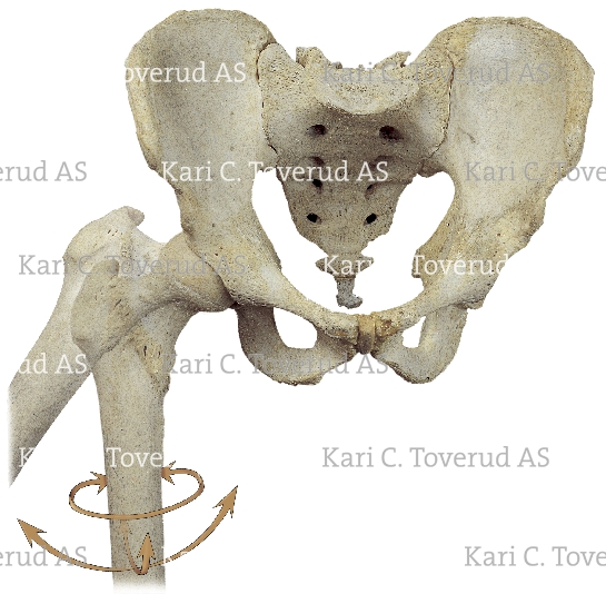
Anterior view of the pelvis showing the ball and socket rotation of the hip joint
Request information on how to purchase usage rights to this image
Please fill out the form below with all the relevant details. If want to request information on more than one illustration, you may prefer to e-mail me directly instead. If you do, please include the link to this illustration and the other illustrations: http://www.karitoverud.com/illustrations/C/749/
 Instagram